During the 10th annual Brain Research Through Advancing Innovative Neurotechnologies ® (BRAIN) Initiative Conference, we announced the winners of the sixth BRAIN Initiative Photo and Video Contest. Each year has produced fascinating depictions of the brain, and this year is no exception. To celebrate ten years of the BRAIN Initiative, we also selected a photo and a video finalist in the special category, BRAIN at 10. Here we feature the winning images and the researchers who created them. Don’t miss your chance to order a free copy of the 2025 BRAIN Initiative calendar.
First Place Video Winner: Expl
oring the 3D nanoarchitecture of neuronal membranes at dendritic spine by labeling Channelrhodopsin-2
The first-place winner in the video category. Credit: Hao-Cheng Gao, Xi Cheng, Alexander Chubykin and Fang Huang, Purdue University.
“Exploring the 3D nanoarchitecture of neuronal membranes at dendritic spines by labeling Channelrhodopsin-2” was made by Hao-Cheng Gao, a PhD student in Fang Huang’s lab at Purdue University. The video provides a high-resolution view of the structure of dendritic spines in thick sections of mouse brain tissue.
Gao’s work involves developing microscopy techniques to achieve single-molecule resolution for studying neuroanatomy of the brain. To create this 3D distribution of ion channels, Gao labeled them with a fluorescent marker and then used a super-resolution imaging technique to capture these tiny structures at a sub-15nm resolution. This approach provides a detailed view of how sensory experiences sculpt neural circuits and synaptic connections.
The imaging technique used in this project bridges the gap between conventional neuroanatomy and ultra-high-resolution analysis, providing exceptional resolution and specificity. The resulting video represents the success of combining the expertise in complementary fields of brain neuron studies, super-resolution imaging, and information visualization.
“[This video] symbolizes the hard work and dedication of everyone involved, highlighting our effort to push the boundaries of BRAIN Initiative research.” – Hao-Cheng Gao, Purdue University
Second Place Video Winner: Spinal motor neurons illuminated by an enhancer AAV
The second-place winner in the video category. Credit: Tanya Daigle and Emily Kussik, Allen Institute for Brain Science
“Spinal motor neurons illuminated by enhancer AAV” was created by Tanya Daigle and Emily Kussik at the Allen Institute for Brain Science. The video shows the detailed structure of motor neurons in a mouse spinal cord progressing from top to bottom.
To generate this video, Dr. Daigle used an enhancer Adeno-Associated Virus (AAV) to drive SYFP2 expression in spinal motor neurons then used a program to stitch together ~2m transverse sections of spinal cord. The resulting video shows a detailed fly-through of the motor neurons along the spinal cord, progressing from the cervical, thoracis, lumbar, and sacral sections.
Dr. Daigle’s research focuses on developing cell type specific viruses for both basic and translational research. Spinal motor neurons selectively deteriorate in many devasting neurodegenerative disorders, such as amyotrophic lateral sclerosis (ALS), and many have no cure or effective therapy. She believes that the enhancer AAV in this video could one day be engineered to deliver a therapeutic cargo to these neurons in humans and improve the health and outcome in various disease states.
“This video represents a significant advancement in our ability to develop viral tools to label cell types within the central nervous system and beyond. We can now visualize diverse cell types within the brain spinal cord, and body, and will soon be able to perturb and monitor them using this approach.” – Tanya Daigle and Emily Kussick, Allen Institute for Brain Science
Third Place Video Winner: Neuronal “fireworks” when the salamander sniffs
The third-place winner in the video category. Credit: Lu Xu, Wenze Li., Eliza Jaeger, Elizabeth Hillman, and Maria Tosches, Columbia University.
“Neuronal ‘fireworks’ when the salamander sniffs” is a collaboration between Lu Xu, Wenze Li, Eliza Jaeger, Elizabeth Hillman, and Maria Tosches at Columbia University. This video illustrates how neurons coordinate their activity to encode different odor identities. According to Dr. Xu, when stimulated by different odors (when the salamander sniffs) the neurons appear like little “fireworks” or notes and chords dancing on a musical score.
To create in vivo recordings of neurons responding to different odors, Dr. Xu immunolabeled neurons to glow green when activated. Odors were presented by puffs of air and images were collected using a technique called swift confocally-aligned planar excitation (SCAPE) microscopy. This novel 3D imaging approach allowed them to observe the activity of a large group of neurons, while also maintaining the resolution needed to observe the detailed structure of each individual neuron.
This video is part of the lab’s ongoing project in deciphering cortical olfactory codes. The salamander brain is much smaller and more accessible than that of mice, making it an ideal model for studying olfactory circuits and neuronal circuits. Furthermore, salamanders offer valuable insights into the evolutionary adaptations related to water-to-land transition in ancestral tetrapods.
“It’s incredible to see how different neurons coordinate to represent odor identity in the salamander brain. They appear like little ‘fireworks’, or notes and chords dancing on a musical score.” – Lu Xu, Columbia University
BRAIN at 10 Video Finalist: Cortical Density
The BRAIN at 10 Video Finalist. Credit: Tyler Sloan, Quorumetrix Studio.
“Cortical Density” was generated by Tyler Sloan at Quorumetrix Studio. The video demonstrates the immense progress that has been made in our ability to visualize the brain. It begins by modeling early Golgi staining in a mouse brain, which typically stains only ~1% of neurons, then progresses to modern methods that allow visualization of 99% of the neurons.
The animation was generated using publicly available data from the Machine Intelligence from Cortical Networks (MICrONS) consortium, a large, concerted effort of scientists from varied backgrounds, who produced massive datasets and turned them into a resource for other researchers. Dr. Sloan used the Connectome Annotation and Versioning Engine (CAVE) computational infrastructure to process a collection of 75,000 neurons. He then simulated the conventional “Golgi-view” by making 99% of those neurons invisible.
This remarkable depiction of the progress in neuron imaging since the discovery of the Golgi stain in 1873 also highlights another form of growth. Historically, much of the history of famous neuroscience discoveries was driven by a few notable individuals or small teams. The MICrONS project represents a fundamental shift of scale, involving massive financial investment and teams of talented scientists from multiple institutions, collaborating to establish and maintain a public resource that is available to the entire field, often before any results are published. To Dr. Sloan, it means that neuroscience has matured into a team sport.
“I hope that my graphics can be helpful for people outside our field to appreciate the elaborate beauty of neural wiring, as well as to boost the intuitions of my fellow neuroscientists.” – Tyler Sloan, Quorumetrix Studio
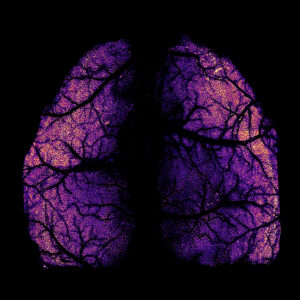
First Place Photo Winner: Whole-cortex scale in vivo two-photon imaging with single-cell resolution in mice

The first-place winner in the video category. Credit: Zongyue Cheng, Jianian Lin, and Meng Cui, Purdue University.
“Whole-cortex scale in vivo two-photon imaging with single-cell resolution in mice” was created by Zongyue Cheng and his colleagues, Jianlan Lin and Meng Cui at Purdue University. The image portrays the entire dorsal neocortex of a mouse brain with remarkable resolution.
Dr. Cheng and his team used the innovative Light Pipe Microscope (LPM) array to capture sequential images at various depths throughout the brain of a transgenic mouse. This approach allowed the team to visualize an entire brain region at once and facilitates comprehensive, large-scale imaging while also achieving single-cell resolution.
This photo represents the sustained dedication and collaborative efforts of the research team over years. It symbolizes the beauty of brain architecture and the breakthrough possibilities that advanced imaging techniques offer in understanding neural dynamics on a large scale.
“This image demonstrates the potential of advanced imaging techniques to capture the dynamics of neurons across the entire brain cortex, symbolizing our pursuit to decode brain mysteries with scientific precision and biological elegance.” – Zongyue Cheng, Purdue University
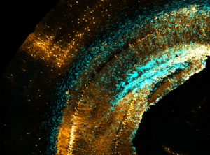
Second Place Photo Winner: Chaos circuitry

The second-place photo winner. Credit: Yusha Sun, University of Pennsylvania
“Chaos circuitry” was produced by Yusha Sun from Hongjun Song’s lab at the University of Pennsylvania. The photograph depicts neurons (orange) that synapse with human glioblastoma tumor cells (blue) in the mouse brain. This image was obtained using a novel method that harnesses the power of trans-synaptic tracing techniques, traditionally used in the circuit neuroscience field, to study the properties of tumor-neuron circuitry.
To generate this image, Sun and team transplanted glioblastoma cells into the brain of an adult mouse and then safely delivered a virus to tumor cells. The virus spread across single synapses from the tumor cells to neighboring neurons. The resulting image reveals striking diversity in the neurons that interact with the tumor cells.
The surprising range of neurons connecting to the tumor cells suggests that glioblastoma may be a brain-wide disease. Sun hopes that this discovery and the improved understanding of tumor-neuron circuitry will help open the door to new ways of fighting brain tumors in people.
“While this is a frightening concept, our improved understanding of tumor-neuron circuitry can open new doors toward fighting this disease.” – Yusha Sun, University of Pennsylvania
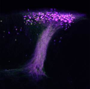
Third Place Photo Winner: Willow on the Island of Calleja

The third-place photo winner. Credit: Lee O. Vaasjo and Maria J. Galazo, Tulane University.
“Willow on the Island of Calleja” by Lee Vaasjo and Maria Galazo at Tulane University depicts a seldom-studied population of neurons, the island of Calleja, that are responsible for receiving complex olfactory information.
The image was generated as part of an effort to study neurons in the frontal areas of the mouse neocortex. Dr. Vaasjo serendipitously labeled the island of Calleja while studying a Syt6-Cre transgenic mouse line. This genetic strategy was combined with a fluorescent reporter and 3D laser scanning fluorescence confocal microscopy was used to capture the anatomy of this population of neurons.
Distinct neuron populations play individual, sometimes indispensable, roles in orchestrating behavior. Drs. Vaasjo and Galazo have investigated this by evaluating the relationship between structure and function at the neuron sub-type level. It is not yet understood how exactly the island of Calleja processes neural signals before relaying them to higher-order brain regions. However, Dr. Vaasjo hopes that their work underscores the importance of paying attention to the serendipitous and unexpected.
“With “Willow on the Island of Calleja,” I sought to represent the benefits of solitary, meditative thought in giving rise to well-founded, flourishing, interconnected ideas. The photographic imagery was inspired by the willow oak trees in Audubon Park in the city of New Orleans. The tree-like arborizations remind me of a reflective stroll in the park.” – Lee O. Vaasjio, Tulane University
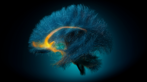
BRAIN at 10 Photo Finalist: Decoding Depression

BRAIN at 10 Photo Finalist. Credit: Christopher Rozell, Mike Halerz, Ki Sueng Choi, and Helen Mayberg, Georgia Institute of Technology, Icahn School of Medicine at Mt. Sinai, and TeraPixel, Inc.
“Decoding Depression” was produced through a collaborative effort by Christopher Rozell, Mike Halerz, Ki Sueng Choi, and Helen Mayberg, at Georgia Institute of Technology, Icahn School of Medicine at Mt. Sinai, and TeraPixel, Inc. The image shows the targeted area in the brain of a patient undergoing deep brain stimulation (DBS) therapy for treatment-resistant depression.
Dr. Rozell and his colleagues implemented an advanced imaging technique called diffusion tensor imaging to capture the brain’s white matter. The 3D reconstruction was done by modeling software. White matter targeted by DBS is highlighted in orange and the remaining structures are highlighted in blue.
According to Dr. Rozell, this image underscores the importance of this work to helping patients while also depicting that the intersection of nature and technology can be intrinsically beautiful. Accurate imaging of white matter is key to successful DBS therapy. This type of imaging is both beautiful and central to developing and implementing new circuit-based therapies.
“This photo represents to us the beauty that arises from the combination of the intricate structure of the brain and the modern technology used to image it.” – Christopher Rozell, Georgia Institute of Technology
Congratulations to all the 2024 winners! Visit the contest page to order a free copy of the 2025 BRAIN Initiative calendar.
Love what you see? Check out more stunning imagery of the brain in previous ‘Art of the BRAIN’ series below:

